Welcome to DERME IMAGE
We specialize in advanced mole mapping using total-body imaging and dermoscopy to support early detection and long-term monitoring of skin changes.
Mole mapping is offered exclusively at Radimed Westmount Square through our collaboration, bringing together advanced imaging and expert dermatologic review.
Together, we deliver a dedicated service designed for early detection and ongoing skin surveillance.

How can
mole mapping
detect early melanoma?
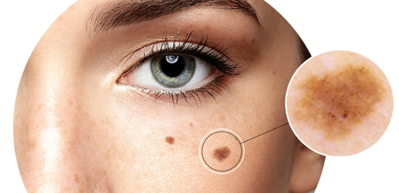
A step-by-step process conducted by expert technicians: a full-body screening is conducted for suspicious moles before a closer examination is done using a dermatoscope (high magnification handheld microscope with polarized light). This technology allows dermatologists to make clinical diagnoses. catching skin cancer in its initial stages.

Track Change. Detect Early.
See What the Eye Can Miss
Mole Mapping Comparison Over Time

Baseline Imaging
-
Full-body polarized photography
-
Dermoscopic lesion documentation
-
Creates a visual reference point
-
Captures lesions that may appear clinically benign today
Follow-Up Comparison
-
Detects subtle interval change
-
Identifies new or evolving lesions
-
Side-by-side dermoscopic comparison
-
Supports earlier melanoma detection

Dermoscopy:
See Beyond the Surface
High-magnification polarized imaging helps reveal structures invisable to the naked eye
What Dermoscopy Detects
Dermoscopy uses polarized magnification to evaluate pigment patterns, vascular structures, and subtle skin changes not visible during a routine visual skin examination.
Why It Matters
Many early melanomas appear clinically subtle. Dermoscopic imaging helps identify suspicious structures earlier and improves long-term lesion monitoring when images are compared over time.
Combined with Mole Mapping
At DERME IMAGE, dermoscopy is integrated with total-body imaging to support both:
-
detection of new lesions
-
monitoring of evolving lesions over time
A New Standard of Care
Dr. Avram Whiteman, Assistant Professor of Family Medicine at McGill University and holder of a Master’s degree in Public Health, has long been passionate about disease prevention. “As the old saying goes, an ounce of prevention is worth a pound of cure,” he says. That philosophy led him to collaborate with Dr. Robert Solomon, a board-certified dermatologist and lecturer at the University of Toronto, to explore the potential of advanced computer-assisted technology designed to detect skin changes earlier and more accurately. They discovered FotoFinder®, a sophisticated digital mole mapping system that captures high-resolution, polarized photographs of the entire body. The technology identifies and records individual skin lesions and stores digital dermoscopic images for future comparison. Unlike a standard skin examination, mole mapping allows dermatologists to log, chart, and document moles across the entire skin surface and track changes over time. This technology is particularly useful for identifying new lesions, including those that may be suspicious for melanoma. The specialized software stores and compares high-resolution images from previous visits, allowing clinicians to detect subtle changes in size, colour, structure, or pattern that might otherwise go unnoticed during routine visual examinations. By documenting the skin at baseline and comparing images at follow-up visits, mole mapping significantly improves the ability to detect melanoma at an earlier stage, when treatment is most effective.

Patient Journey
Last year, Julie noticed a spot on her arm that made her uneasy. Having experienced a few sunburns over the years, she worried if it could be something serious. After consulting her family doctor, she was referred to a dermatologist - but the wait time for an appointment was more than six months. Concerned about delaying the evaluation, Julie looked for alternatives. A friend recommended mole mapping with DERME IMAGE at Radimed Westmount Square. Within two days, Julie underwent a full-body mole mapping exam. The spot on her arm turned out to be benign, but the exam revealed a smaller spot on her back that raised concern. A biopsy revealed the lesion was a malignant melanoma - but fortunately, it was caught early. It was removed, and the excision proved curative. Like other routine health exams, Julie will continue to return for annual preventive screenings.

Who Should Consider It?
Skin cancer is the most common cancer. A quick, non-invasive skin screening can give you peace of mind, especially if you have risk factors for melanoma.
Do any of these apply to you?
-
You are over the age of 16.
-
You have many moles (more than ~50).
-
There’s a family history of skin cancer.
-
You’ve had melanoma before.
-
You have very large or atypical moles (several centimeters across).
-
You’ve noticed changes in a mole’s size, shape, color, or symptoms (itching, bleeding).
-
You’ve noticed new moles.
-
You had severe, blistering sunburns in childhood or adolescence.
-
You have fair or sensitive skin that burns easily.
-
You are immunocompromised.
If you answered “yes” to any of the above, consider booking a skin screening.
Note: If a spot is rapidly changing, bleeding, or looks very different from your other moles (“the ugly duckling”), seek assessment promptly.